
Figure 2.
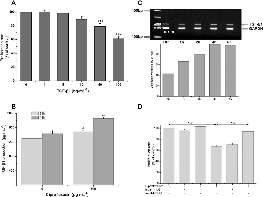
Ciprofloxacin-induced cell cycle arrest is mediated through TGF-β1. (A) MTT assessed proliferation of HT-29 cells after treatment with 0–100 ng·mL−1 of TGF-β1 for 3 days. (B) TGF-β1 production by HT-29 cells after 24 and 48 h treatment with vehicle (0) or ciprofloxacin (100 µg·mL−1) evaluated by elisa in cell culture supernatants. (C) Expression of TGF-β1 mRNA by HT-29 cells. Total RNA was extracted from HT-29 cells after treatment with ciprofloxacin (100 µg·mL−1) for 1 to 6 h and multiplex RT-PCR for TGF-β1 and GAPDH was performed. Upper panel: representative PCR blot of mRNA expression for TGF-β1 and GAPDH. Lower panel: the ratio of the integrated density of TGF-β1 divided by that of GAPDH, measured by densitometry analysis, is expressed as a percentage of the maximum TGF-β1 mRNA expression after stimulation with ciprofloxacin (100 µg·mL−1). One experiment representative of three independent experiments with similar results is shown. (D) MTT assessed proliferation of HT-29 cells after treatment with ciprofloxacin (100 µg·mL−1) for 6 days in the presence of 10 µg·mL−1 of neutralizing anti-human-TGF-β1 (LAP) antibody or 10 µg·mL−1 of purified mouse IgG2a isotype control (*P < 0.05, **P < 0.01, ***P < 0.001). GAPDH, glyceraldehyde 3-phosphate dehydrogenase; MTT, 3-(4,5-dimethylthiazol-2-yl)-2,5-diphenol tetrazolium bromide; RT-PCR, reverse transcription polymerase chain reaction; TGF, transforming growth factor.