
Figure 5.
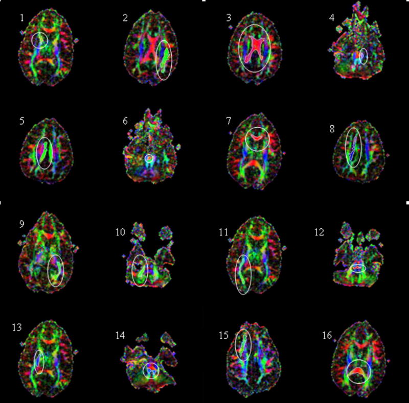
ROQS segmentation of various structures in the brain displayed on axial directionally encoded color FA images. The ability of ROQS to trace a variety of structures demonstrates its robustness. ROIs shown in white within each white ellipse are 1) Anterior limb of internal capsule; 2) Arcuate Fasciculus; 3) Body of the Corpus Callosum; 4) Cerebral Peduncle; 5) Cingulum; 6) Decussation of Superior Cerebellar Peduncle; 7) Genu of Corpus Callosum; 8) High Centrum Semiovale; 9) Optic Radiations; 10) Middle Cerebellar Peduncle; 11) Inferior Longitudinal Fasciculus; 12) Pontine Crossing Tract; 13) Posterior Limb of Internal Capsule; 14) Superior Cerebellar Peduncle; 15) Superior Longitudinal Fasciculus; 16) Splenium of Corpus Callosum