
Figure 3.
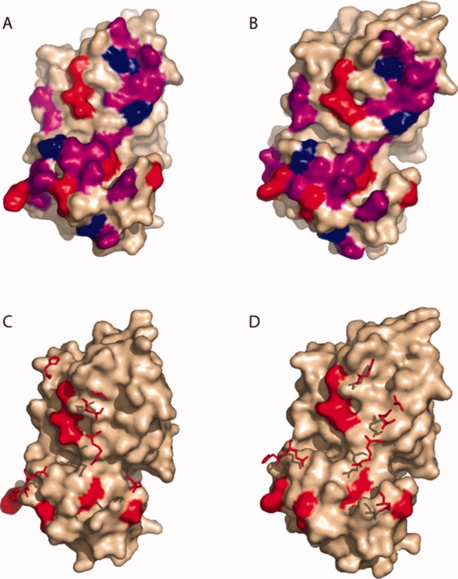
Positions of mutated residues. Mutated residues are colored on the dimer interface of the monomer structures. In panels (A) and (B), Class I (hot spot) residues are colored red. Class II residues are purple and Class III residues are blue in the reduced and oxidized forms, respectively. Panels (C) and (D) show how the hot spot residues on one monomer are positioned relative to hot spots across the interface. The surface of one monomer is shown (wheat colored), with the seven Class I positions highlighted red. The seven Class I positions from the other monomer are shown as stick structures. (C) Reduced Form. (D) Oxidized form.