
Figure 5.
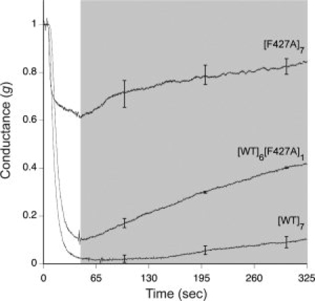
Normalized macroscopic channel current traces monitoring dissociation of LFN-streptavidin from F427A-containing mutants during perfusion of the cis compartment. Streptavidin was bound to LFN through a C-terminal biotin tag on the latter. LFN-streptavidin (10 nM) was added at time zero to channels formed by [WT]7, [F427A]7, or [WT]6[F427A]1. After channel blockage approached its maximum value, we perfused the cis compartment and monitored current while holding the membrane potential at Δψ = +5 mV. The shaded area shows the period during which perfusion (∼ 7 mL/min) was conducted. LFN-streptavidin dissociation is manifested by the rise in current during perfusion. The data plotted are averages of three experiments, with standard error shown for every 1000 data points.