
Figure 3.
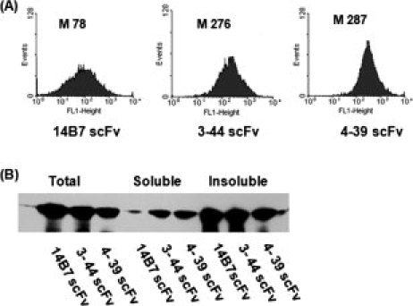
Analysis of isolated scFv clones. (A) FACS histograms of MCA cells expressing 14B7*, 3-44, or 4-39 scFvs. (B) Solubility of the scFvs following expression in the bacterial cytoplasm. Cells were grown at 25°C, and protein synthesis was induced with 1 mM IPTG for 4 h and harvested. Proteins were resolved by SDS-PAGE and scFvs were detected by Western blotting using anti-His-HRP at a 1:10,000 dilution. Samples were normalized by loading aliquots equivalent to OD600 = 2.