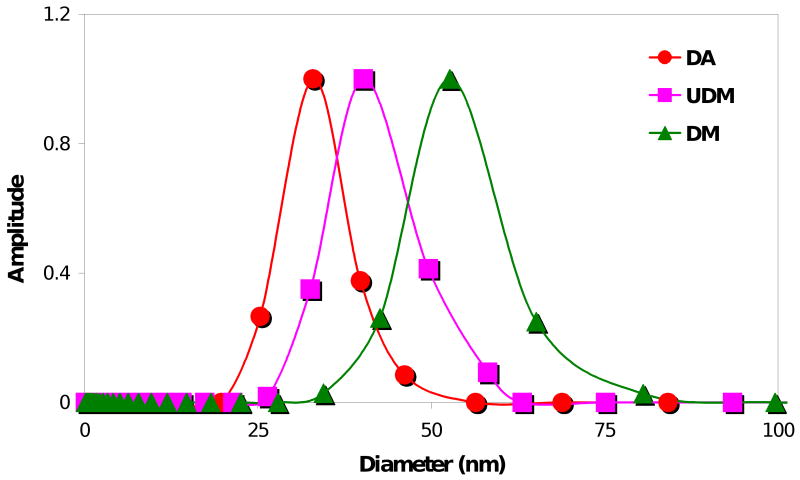
Figure 6.
Characterization of the micellar sizes by DLS for the three macromonomers in deionized water at a concentration of 5 mg/mL. The 4KG5 DA, UDM, and DM micelles were found to have diameters of 33.5 ± 1.4 nm, 40.3 ± 1.6 nm, and 56.3 ± 2.3 nm, respectively. The sizes were calculated by volume measurement and expressed as Dav ± S (average diameter ± standard deviation).

