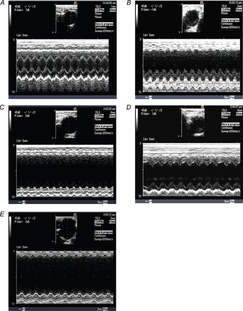
Figure 2. Representative echocardiograms showing left ventricular chamber dilatation and dysfunction after MI (B and C) as well as the effect of 17β-oestradiol (0.42 and 42 μg day−1) on left ventricular dilatation and dysfunction (D and E) in comparison with a normal mouse heart (A).
A, sham OVX + sham MI; B, sham OVX + MI + placebo; C, OVX + MI + placebo; D, OVX + MI + E2 (0.42 μg day−1); and E, OVX + MI + E2 (4.2 μg day−1).

