
Figure 4. Electron microscopy of transfected cells.
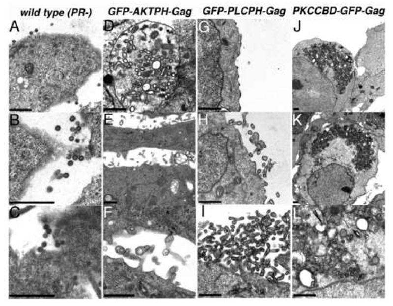
Cells were transfected with constructs expressing protease-minus but otherwise wild type HIV Gag (wild type Pr-; panels A-C), GFP-AKTPH-Gag (panels D-F), GFP-PLCPH-Gag (panels G-I), or PKCCBD-GFP-Gag (panels J-L). At 3 days post-transfection, cells were fixed, post-fixed, dehydrated, embedded, sectioned, and stained for electron microscopy as described in the Materials and Methods. As illustrated, relatively homogeneous wild type PR- particles were observed at the surfaces of cells (A-C), while structures assembled by GFP-AKTPH-Gag proteins were pleiomorphic and appeared as VLPs or darkly rimmed vesicles within intracellular vacuoles (D), particles released at the cell surface (E), or preformed VLPs near the cell surface or within attached or sheared filopodia (F). In contrast, assembly of spherical or tubular GFP-PLCPH-Gag VLPs occurred specifically at cell surfaces (G-I), and PKCCBD-GFP-Gag proteins assembled into large intracellular aggresomes (J-L).