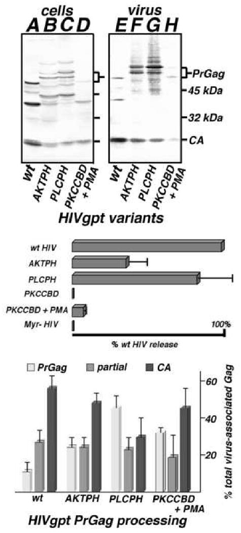
Figure 6. Virus-like particle release and processing.

Top panel. Cell lysate (A-D) and VLP (E-H) samples were collected from cells transfected with wt HIVgpt (wt), HIVgpt-AKTPH (AKTPH), HIVgpt-PLCPH (PLCPH) or HIVgpt-PKCCBD (PKCCBD), and either untreated or treated with 1 μM PMA (+PMA). Gag proteins in samples were fractionated by SDS-PAGE, and detected by immunoblotting using an anti-HIVCA primary antibody. Size marker mobilities, as well as PrGag and CA bands, are indicated. Middle panel. Virus-like particle assembly and release levels, normalized to that of wt HIVgpt, were determined as described in Figure 2, including a myristoylation-minus HIVgpt construct (Myr- HIV) as a negative control. Values derive from two (PKCCBD, Myr- HIV), three (PKCCBD), or four (AKTPH, PKCCBD + PMA) independent experiments, with standard deviations as shown. Bottom panel. PrGag processing levels for VLPs produced from cells expressing wt HIVgpt (wt), AKTPH, PLCPH, and PKCCBD (in the presence of PMA) were determined by densitometric quantitation of PrGag (light gray), processing intermediates (medium gray), and CA bands (dark gray) from immunoblots. Values represent percentages of the total virus-associated Gag levels (plus standard deviations) and derive from three (PKCCBD +PMA), four (AKTPH, PLCPH), or seven (wt) independent VLP preparations.
