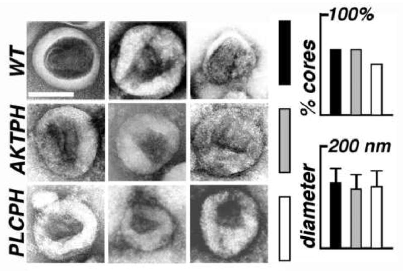
Figure 7. Electron microscopy of virus-like particles.

Wild type (wt; black bars) HIVgpt, HIVgpt-AKTPH (AKTPH; gray bars), and HIVgpt-PLCPH (PLCPH; white bars) virus-like particles (VLPs) were lifted onto EM grids, stained, dried, and imaged by electron microscopy (EM). Micrographs show VLPs with central, roughly conical cores, and the white size bar in the upper right panel corresponds to 100 nm for all pictures. The graphs on the right shows the average VLP diameters, and the percentages of VLPs with discernable, roughly conical or cylindrical cores: each value was derived from 100 separate VLP images.
