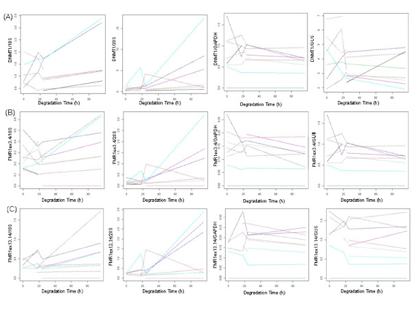
Figure 3.
Normalization of FMR1 and DNMT1 mRNA to 18S, 28S, GAPDH and GUS, expressed as a function of degradation time. Each sample (listed in Table 1) is represented by a differently colored line whose number varied between 8 and 14 for the nine plots; depending on completeness of the data set for the variables examined. (A) DNMT1 mRNA normalized to 18S (p = 0.066), 28S (p = 0.066), GAPDH (p = 0.388), GUS (p = 0.774); (B) FMR1ex3.4 mRNA normalized 18S (p = 0.11), 28S (p = 0.11), GAPDH (p = 0.51), GUS (p = 0.254); (C) FMR1ex13.14 mRNA normalized 18S (p = 0.11), 28S (p = 0.11), GAPDH (p = 0.11), GUS (p = 0.34).

