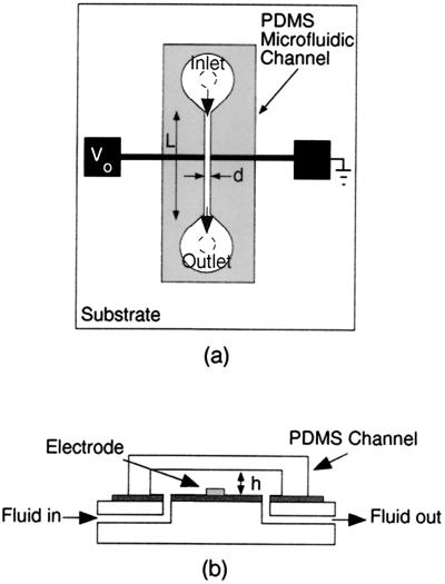
Figure 1.
Schematic illustration of the integrated microfluidic device. (a) Top view shows the entire device, including electrode configuration, inlet and outlet holes for fluid, and the PDMS microfluidic channel. The electrodes are made of gold and are 50 μm wide. The distance, d, separating the electrodes is 30 μm. The width of the PDMS microfluidic channel is also d, the length, L, is 5 mm, and the height, h, is either 30 μm or 40 μm. (b) Side view along the vertical axis of the device shows a detailed view of fluid delivery. Fluid delivery is accomplished with a syringe pump at nonpulsatile rates ranging from 1 to 300 μl/hr.

