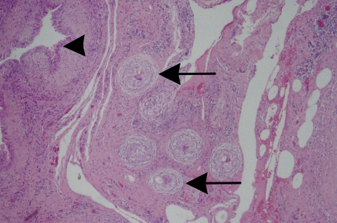
FIG. 7.
The excised pleura from a patient with North American paragonimiasis demonstrates mesothelial hyperplasia (arrowhead), an acute and chronic inflammatory cell infiltrate with eosinophils, and eggs of P. kellicotti that are entrapped in nonnecrotizing granulomas that are beginning to be surrounded by concentric fibrosis (arrows). Hematoxylin and eosin staining was used. Magnification, ×40.

