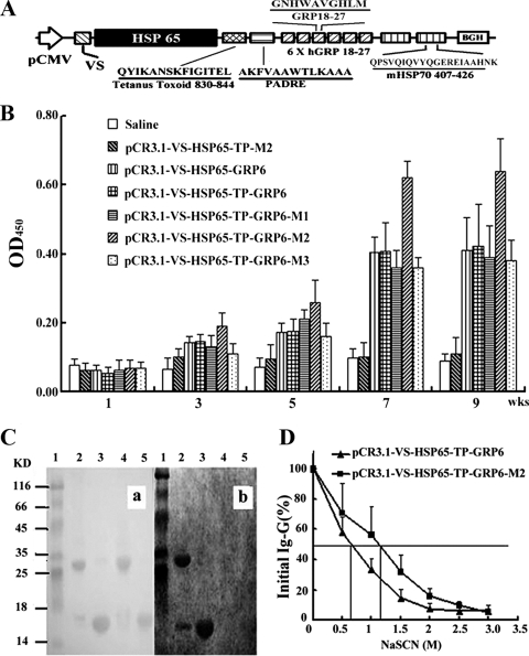
FIG. 1.
Characterization of GRP-specific IgG from immunized mice. (A) Schematic diagram of pCR3.1-VS-HSP65-TP-GRP6-M2. In this DNA vaccine, the VS cDNA (VS) was placed under the control of promoter pCMV (arrow), followed sequentially by the genes encoding for HSP65 (black box), tetanus toxoid 830-844 (T), PADRE (P), six tandem repeats of human GRP18-27 (6 X hGRP 18-27; GRP6), and two copies of mycobacterial HSP70407-426 (mHSP70 407-426; M2). (B) Mice immunized with pCR3.1-VS-HSP65-TP-GRP6-M2 produced the highest titers of anti-GRP antibody than any other groups, especially at 7 weeks (wks) after the initial immunization (P < 0.001). OD450, OD at 450 nm; KD, kilodaltons. (C) Specificity of anti-GRP antibodies was verified using immunoblot analysis. Proteins transferred onto nitrocellulose membranes were stained with Ponceau red (a) or incubated with sera from immunized mice (b). Lanes 1, protein markers; lanes 2, rhVEGF121-GRP18-27 without DTT; lanes 3, rhVEGF121-GRP18-27 with DTT; lanes 4, rhVEGF121 without DTT; lanes 5, rhVEGF121 with DTT. (D) Anti-GRP antibodies from sera of mice immunized with HSP70-containing plasmids had relative avidity significantly (P < 0.0001) higher than that for the group without, according to modified ELISA.

