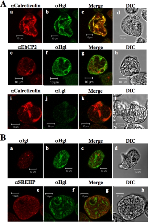
FIG. 10.
Subcellular localization of secretory proteins after treatment of wild-type cells with brefeldin A. Cells were treated with 100 μg/ml BFA for 3 h, after which they were fixed and stained with antibodies specific for a variety of secretory proteins. (A) BFA treatment phenocopies expression of EhRabAQ84L with respect to the localization of Hgl (b and f), calreticulin (a and i), Lgl (j), and EhCP2 (e). In treated cells these proteins exhibit near-complete colocalization in intracellular compartments, some of which are perinuclear. (B) Similar to EhRabAQ84L overexpression, BFA does not alter the localization of Igl or SREHP. Colocalized antigens are shown in yellow (A, panels c, g, and k, and B, panels c and g) and corresponding differential interference contrast images (A, panels d, h, and l, and B, panels d and h) are provided. Bars, 10 μm.

