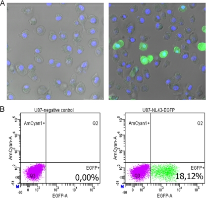
FIG. 2.
Discrimination between infected and noninfected cells. U87.CD4.CCR5.CXCR4 cells were infected with HIV-1-NL4.3-EGFP (right) or were mock infected (left). Twenty-four hours later, cells were washed with PBS, trypsinized, and fixed in 2% paraformaldehyde for 10 min. (A) Afterwards, nuclei were stained for 5 min with DAPI (4′,6-diamidino-2-phenylindole). Subsequently, cells were washed three times with PBS, resuspended in PBS, and visualized by fluorescence microscopy. (B) After fixation, cells were analyzed by flow cytometry.

