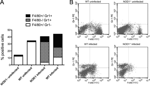
FIG. 3.
NOD1 affects L. monocytogenes-induced recruitment of granulocytes and resident and inflammatory monocytes to the inflammatory site. NOD1−/− and WT peritoneal exudate cells were harvested from mice before (five WT mice and four NOD1−/− mice) or 2 days after (6 WT mice and six NOD1−/− mice) i.p. infection with 105 L. monocytogenes CFU. The mean percentages of cells that were stained with anti-Gr1 and/or anti-F4/80 to differentiate between polymorphonuclear leukocytes and inflammatory monocytes and resting macrophages and monocytes in the peritoneal exudates from individual mice are shown. Differences between the total numbers of harvested peritoneal cells from L. monocytogenes-infected and uninfected NOD1−/− and WT animals are not significant. Differences between the percentages of F4/80+ Gr1− cells in infected and uninfected peritoneal exudates from WT or NOD1−/− mice are significant (P < 0.001, Student's t test). Differences between the percentages of F4/80+ Gr1− cells in peritoneal exudates from infected or uninfected WT and NOD1−/− mice are significant (P < 0.001, Student's t test). Differences between the percentages of Gr1+ F4/80− cells from infected WT and NOD1−/− mice are significant (P < 0.001, Student's t test). Differences between the percentages of Gr1+ F4/80+ peritoneal exudate cells from WT and NOD1−/− mice are significant (P < 0.02, Student's t test). The results of one of two independent experiments are shown. (B) Levels of expression of F4/80+ and Gr1+ cells in the peritoneal exudates of representative mice before or after infection with L. monocytogenes. FITC, fluorescein isothiocyanate; PE, phycoerythrin.

