
FIG. 3.
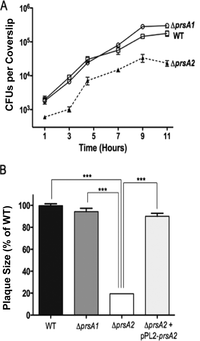
The ΔprsA2::erm mutant is capable of intracellular growth but is defective for cell-to-cell spread in tissue culture cells. (A) J774 macrophage-like cells were infected with the WT or the ΔprsA1 or ΔprsA2::erm mutant at an MOI of 0.1. After 30 min, the monolayers were washed and gentamicin (50 μg/ml) was added at 1 h postinfection. The data shown are representative of at least three independent experiments done in duplicate. □, wild type; ○, prsA1; ▴, ΔprsA2::erm. (B) Plaque formation in mouse L2 fibroblast cells. Monolayers of L2 cells were infected with the WT or ΔprsA1, ΔprsA2::erm, or ΔprsA2::erm + pPL2-prsA2 mutant for 1 h and washed with PBS, and 20 μg/ml gentamicin was added. At least 15 plaques were measured in three independent experiments for all strains; measurements represent plaque size comparisons with respect to the WT (set at 100%). ***, statistically significant value (P < 0.0001) as calculated using a one-way way analysis of variance with Tukey's multiple comparison test (GraphPad v.6.0A).