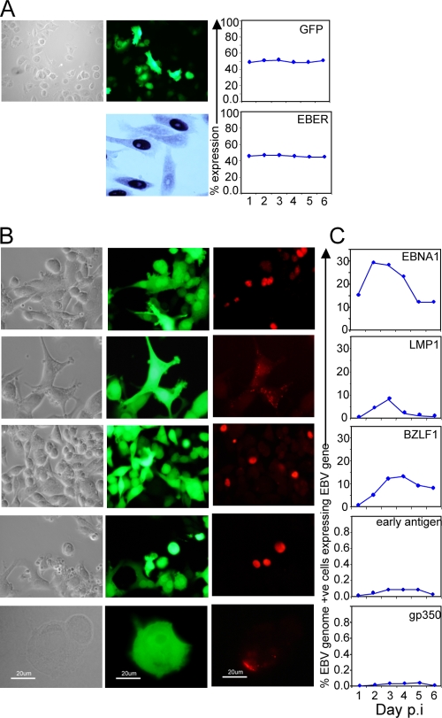
FIG. 3.
Viral gene expression at the single epithelial cell level. (A) Photomicrographs of phase-contrast, GFP fluorescence (upper panels) and EBER ISH (lower panel) of AdAH cultures at 72 h postinfection. GFP and EBER expression were quantified and are represented as the percent expression for an average experiment up to 6 days postinfection. (B) Photomicrographs of phase-contrast and GFP fluorescence of AdAH cells sorted for GFP expression 24 h postinfection. Cells were stained for expression of the latent gene products EBNA1 and LMP1 plus the lytic gene products BZLF1, early antigen EA, and the late antigen gp350. (C) Expression of the latent and lytic gene products was quantitated daily to 6 days postinfection from a population of 100% infected (GFP +ve) cells.

