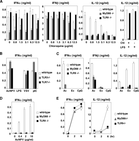
FIG. 1.
Involvement of the TLR pathway in the production of type I IFN by AcNPV in immune cells and in mice. (A) PECs (2 × 10⁵ cells/well) prepared from wild-type, MyD88-deficient, or TLR9-deficient mice were stimulated with AcNPV (10 μg/ml) at the indicated concentrations of chloroquine. These cells were also treated with LPS (10 μg/ml) in the presence (+) or absence (−) of chloroquine (Chl) (12.5 μg/ml) (right). After 24 h of incubation, the production of IFN-α, IFN-β, and IL-12 in culture supernatants was determined by ELISA. (B) PECs (2 × 10⁵ cells/well) prepared from wild-type, TLR3-deficient, or TLR7-deficient mice were stimulated with AcNPV (10 μg/ml), LPS (10 μg/ml), VSV (NCP mutant, MOI of 0.1), or poly(I:C) (pIC) (50 μg/ml). After 24 h of incubation, production of IFN-α in culture supernatants was determined by ELISA. (C) PECs prepared as described for panel A were transfected with AcNPV DNA (Ac) (25 μg/ml), E. coli DNA (Ec) (25 μg/ml), or mCpG (CpG) (1 μg/ml). After 24 h of incubation, production of IFN-α, IFN-β, and IL-12 in the culture supernatants was determined by ELISA. (D) Splenic pDCs (2 × 10⁵ cells/well) prepared from wild-type, MyD88-deficient, or TLR9-deficient mice were stimulated with AcNPV (10 μg/ml). After 24 h of incubation, production of IFN-α in the culture supernatants was determined by ELISA. (E) AcNPV (100 μg/mouse) was intraperitoneally inoculated into wild-type, MyD88-deficient, or TLR9-deficient mice, and levels of IFN-α and IL-12 production in sera were determined by ELISA at the indicated time points. Data are shown as the means ± standard deviations.

