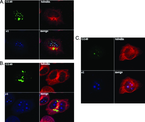
FIG. 3.
The M140 protein colocalizes with viral replication compartments and adjacent to condensed MTOCs in infected macrophages. (A and B) IC-21 macrophages were transfected with a plasmid expressing a GFP-M140 fusion protein under the control of the M140 promoter. The transfected cells were infected with WT MCMV 48 h later and processed for confocal microscopy at 24 (A) or 48 (B) h postinfection. Mouse anti-tubulin and rabbit anti-e1 antibodies were used, followed by goat anti-mouse-tetramethyl rhodamine isocyanate or -fluorescein isothiocyanate and Alexa Fluor 647 goat anti-rabbit conjugates before analysis on a Zeiss 510 confocal microscope. Results were similar when transfected cells were superinfected with RVΔ140 instead of WT virus. (C) NIH 3T3 fibroblasts were transfected and infected as described above for 12 h before processing for confocal microscopy using the antibodies described above.

