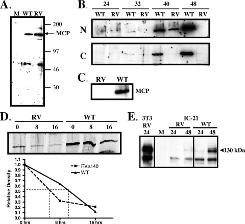
FIG. 4.
Steady-state levels of the MCP and a tegument protein are reduced in RVΔ140-infected macrophages. (A) Confirmation of the specificity of the rabbit antiserum. Two million NIH 3T3 fibroblasts were infected with RVΔ140 (RV) or revertant virus (WT) at an MOI of 2 or were mock (M) infected. Cell lysates were harvested at 24 h postinfection and subjected to Western blot analysis using rabbit antiserum raised against an M86 peptide. (B) Steady-state levels of MCP in nuclear (N) and cytoplasmic (C) fractions of RVΔ140 (RV) and revertant (WT) virus-infected macrophages. IC-21 macrophages were infected at an MOI of 4 with sucrose gradient-purified viruses. Cell lysates were harvested at the indicated hours postinfection, and fractionation was performed using the NE-PER kit. Western blotting utilized the antisera described above. (C) Steady-state levels of MCP in infected primary macrophages. Peritoneal exudate cells from BALB/c mice were infected with sucrose gradient purified revertant virus (WT) or RVΔ140 (RV) at an MOI of 5. Cell lysates were harvested at 48 h postinfection and processed for Western blot analysis using antisera to the MCP as described above. (D) Protein synthesis block to assess stability of MCP expression in mutant and revertant virus-infected macrophages. IC-21 macrophages infected as described above were treated with 100 μM anisomycin starting at 40 h postinfection. Total cell lysates were harvested at the indicated times posttreatment (hours) and subjected to Western blot analysis for the MCP and actin (not shown). Protein bands were quantitated using the Odyssey infrared imaging system, normalized to actin levels, and graphed as a function of time postblock. (E) Levels of tegument protein M25 are reduced in mutant virus-infected IC-21 macrophages. NIH 3T3 fibroblasts (3T3) or IC-21 macrophages were mock infected (M) or infected at an MOI of 2 with revertant (WT) or mutant (RV) virus. Total cell lysates were harvested at the indicated hours postinfection for Western blot analysis using a monoclonal antibody specific for MCMV tegument protein M25.

