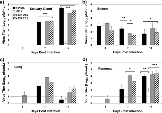
FIG. 1.
Virus titers of wild-type viruses and ΔM33BT2 in the salivary glands (a), spleen (b), lungs (c), and pancreas (d) following i.p. inoculation. Five-week-old BALB/c mice were inoculated i.p. with 2 × 10⁶ PFU of K181 (K-Perth), ΔM33BT2 (ΔM33), M33R D1.6, and M33R F2.1. Titers of tissue sonicates were determined at 3, 7, and 14 dpi by plaque assay on NIH 3T3 cells. The mean virus titer (log10 PFU/ml) and standard error are shown for K181, ΔM33, and M33R F2.1 (n = 12) for a total of three separate experiments. The M33R D1.6 virus was included in one study and is presented as mean virus titer and standard deviation (n = 4). The limit of detection for the plaque assay is shown by the dotted line. Virus titers depicted below the limit of detection indicate where samples were negative at the limit of detection (e.g., 10−1 dilution) but positive for virus in undiluted samples. The P value is shown in the diagram: *, P < 0.05; **, P < 0.01; ***, P < 0.001.

