Abstract
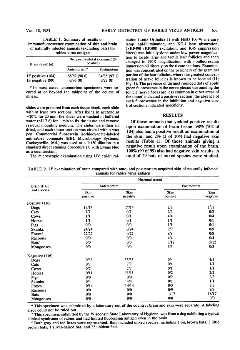
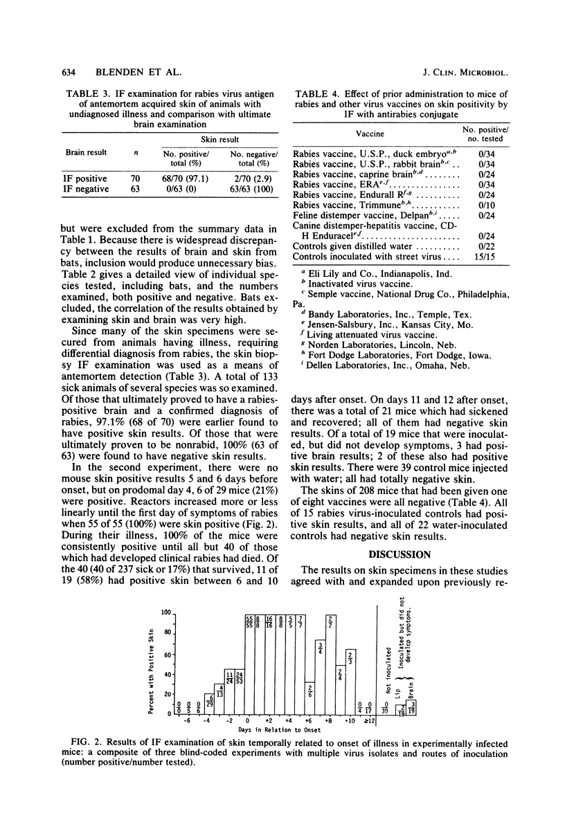
Correlations were made on immunofluorescence positivity to antirabies conjugate between cranium-derived nerve fibers in skin and traditional samplings of brain tissue from several species and illness categories of animals with naturally acquired rabies. The overall correlation of results from all categories was about 98% (n, 104) for those that were brain positive and 100% (n, 99) for those that were brain negative. Some animals that ultimately developed rabies were found to have immunofluorescence-positive results 2 or more days before the onset of clinical signs in both natural and experimental infections. The percentage of those with positive skin immunofluorescence results increased as the onset of symptoms approached. From the midcourse period of illness to death, the correlation between skin and brain approached 100%. Different vaccines, commonly given to prevent rabies and other diseases of dogs and cats, were administered to groups of mice and were found to not produce false-positive results when their skin was examined by immunofluorescence for rabies virus antigen. These data suggest that examination of surgical biopsy specimens by immunofluorescence for rabies virus antigen is a useful and reliable diagnostic tool to evaluate the rabies status of biting dogs or cats, or to confirm a clinical diagnosis of rabies in the species tested. The biopsy evaluation of any other species as a means of assessing bite risk is not suggested by these data.
Full text
PDF
Selected References
These references are in PubMed. This may not be the complete list of references from this article.
- Blenden D. C., Frost J. W., Wachendörfer G., Dorsey C. Identification of rabies virus antigen in the skin of foxes. Zentralbl Veterinarmed B. 1980;27(9-10):698–704. doi: 10.1111/j.1439-0450.1980.tb02024.x. [DOI] [PubMed] [Google Scholar]
- Blenden D. C. Rabies in a litter of skunks predicted and diagnosed by skin biopsy. J Am Vet Med Assoc. 1981 Oct 15;179(8):789–791. [PubMed] [Google Scholar]
- Bryceson A. D., Greenwood B. M., Warrell D. A., Davidson N. M., Pope H. M., Lawrie J. H., Barnes H. J., Bailie W. E., Wilcox G. E. Demonstration during life of rabies antigen in humans. J Infect Dis. 1975 Jan;131(1):71–74. doi: 10.1093/infdis/131.1.71. [DOI] [PubMed] [Google Scholar]
- Lodmell D. L., Bell J. F., Moore G. J., Raymond G. H. Comparative study of abortive and nonabortive rabies in mice. J Infect Dis. 1969 Jun;119(6):569–580. doi: 10.1093/infdis/119.6.569. [DOI] [PubMed] [Google Scholar]
- Moseley B. L., Morehouse L. G., Kintner L. D. Responses in dogs exposed to attenuated rabies virus. Am J Vet Res. 1968 Nov;29(11):2113–2118. [PubMed] [Google Scholar]
- Smith W. B., Blenden D. C., Fuh T. H., Hiler L. Diagnosis of rabies by immunofluorescent staining of frozen sections of skin. J Am Vet Med Assoc. 1972 Dec 1;161(11):1495–1501. [PubMed] [Google Scholar]
- Umoh J. U., Blenden D. C. The dissemination of rabies virus into cranial nerves and other tissues of experimentally infected goats and dogs and naturally infected skunks. Int J Zoonoses. 1982 Jun;9(1):1–11. [PubMed] [Google Scholar]


