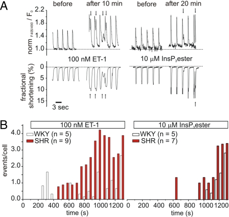
Fig. 4.
Analysis of extra-systolic Ca2+ release events in indo-1 AM-loaded ventricular myocytes during hypertrophy. (A) Representative traces for global Ca2+ transients and cellular contraction recorded from SHR myocytes before and after stimulation with 100 nM ET-1 or 10 μM IP3 ester. Arrows indicate extra-systolic events. (B) Extra-systolic Ca2+ release events per cell during stimulation with 100 nM ET-1 or 10 μM InsP3 ester.

