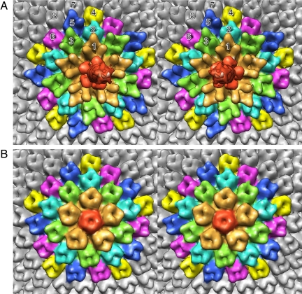
Fig. 2.
Stereodiagrams of the pentasymmetrons at (A) the unique vertex and (B) the 5-fold axis opposite the unique vertex. Note the difference in the density distribution of the pentameric capsomer (red) in A and B. The top end of the narrow spike can also be seen in A. Although all of the major capsomers (Vp54 trimers) surrounding the pentasymmetrons and in the pentasymmetron shown in B have very similar density distributions, this is not the case for the pentasymmetron at the special vertex shown in A. The peripentonal capsomers around the unique vertex are shown in gold and have an extended amino acid insertion. The 10 capsomers surrounding the peripentonal capsomers are colored blue and green. Whereas the green capsomers are similar to the peripentonal capsomers, the blue capsomers are similar to the major capsomers. The capsomers in the outer pentasymmetron ring are colored yellow, dark blue, and pink. The yellow and pink capsomers are similar in structure to the major capsid protein, whereas the dark blue capsomers have a slightly different density distribution.

