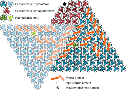
Fig. 4.
Diagrammatic organization of capsomers as seen from inside the virus viewed toward the unique vertex. Within each quasihexagonal capsomer, there are 3 Vp54 monomers, each represented by a dot (blue within one trisymmetron, light blue within the neighboring trisymmetron, and red within the pentasymmetron). Some of the capsomers are associated with minor capsid finger proteins, shown in orange in one trisymmetron and a lighter orange in the neighboring trisymmetron.

