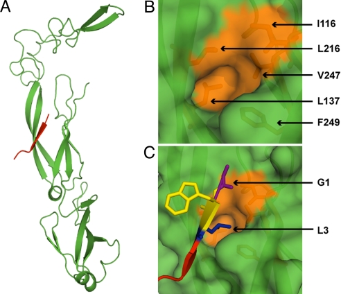
Fig. 1.
Peptide-binding site on Ydj1. (A) The structure (PDB ID: 1NLT) of the peptide binding fragment of Ydj1p (green) in complex with a peptide substrate GWLYEIS (red). (B) Surface representation of the active pocket on Ydj1p. Residues forming the hydrophobic pocket are colored orange and shown in stick representation. The peptide fragment is removed for a clear view of the binding pocket. (C) Orientation of peptide substrate near the active site. The side chain of L3 fits the active pocket. Residue numbering on the peptide conforms to the description in Results.

