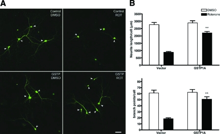
Figure 3.
GSTP1 protected primary cortical cultures from rotenone-induced neurotoxicity. A: Representative images of mouse primary cortical neuron-enriched cultures overexpressing GFP only (control) or human GSTP1A together with GFP (GSTP) and treated with 2 nmol/L rotenone or DMSO as control for 2 days. Arrowheads indicate the MAP2+ neurons. Scale bar = 50 μm. B: Neurite length and complexity on GFP+/MAP2+ neurons were measured using the MicroBrightField Neurolucida software (**P < 0.01 compared with rotenone-treated vector control; n = 55 to 70 cells from ∼20 random selected fields and two independent cultures).

