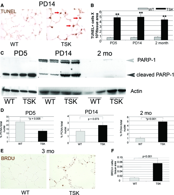
Figure 2.
Prominent apoptosis with reduced proliferation in the neonatal TSK airspace. A: TUNEL staining of the PD14 wild-type and TSK lung shows enhanced epithelial cell death in the TSK airspace. Arrows denote sites of positive staining in the TSK lung. Magnification = original ×20. B: Quantitative immunohistochemistry shows maximal cell death in the PD5 TSK lung that decreases with time. C: Western blotting for PARP cleavage products in lysates from wild-type and TSK lungs at PD5, PD14, and 2 months of age. Increased PARP cleavage occurs during neonatal development in the TSK lung. Gray arrowhead indicates intact PARP-1 protein. Black arrowhead denotes PARP-1 cleavage product. Actin was used as a loading control. D: Quantitative immunohistochemistry of proliferating cell nuclear antigen staining normalized to total cell count in wild-type and TSK airspace at PD5, PD14, and 2 months of age. The mitotic index is reduced in the PD5 TSK airspace but increased by adulthood. E: Representative BRDu staining in adult wild-type versus TSK lungs. Increased BRDu labeling is observed in the TSK airspace. Magnification = original ×20. F: Quantitative immunohistochemistry of BRDu labeling confirms increased staining in the TSK lung. *P < 0.05, **P < 0.001.

