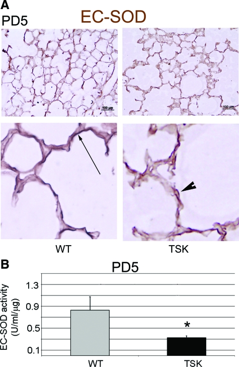
Figure 8.
Abnormal EC-SOD deposition and activity in the TSK lung. A: Representative immunohistochemical staining of EC-SOD in the PD5 wild-type versus TSK lung. Top panel: Low power image demonstrating intact linear staining along alveolar walls in wild-type lungs but marked discontinuity and fragmentation in the TSK lung. Scale bar = 100 μm. Bottom panel: High power view emphasizes fragmentation of EC-SOD in the TSK lung. Arrow depicts continuous EC-SOD staining in the wild-type lung. Arrowhead shows fragmented staining pattern in TSK lung. Magnification = original ×100. B: EC-SOD activity in PD5 lung lysates. Reduced EC-SOD activity is detected in PD5 TSK lung compared with age-matched wild-type lungs. *P < 0.05.

