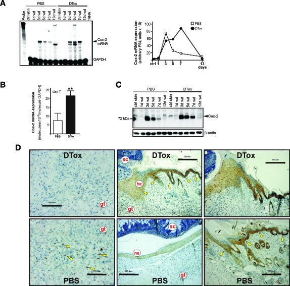
Figure 10.
Cox-2 expression in macrophage-depleted wounds. A: Cox-2 mRNA expression during skin repair in PBS- and DTox-injected mice as indicated (left panel). A quantification of Cox-2 mRNA from data points of the respective RNase protection assay gel is shown in the right panel. B: qRT-PCR quantifications of Cox-2 mRNA expression from 7-day wound tissue from 5 individual mice (n = 5) to demonstrate the individual variability of the respective signal. Bars indicate the mean ± SD obtained from wounds (n = 3) isolated from five individual animals (n = 5). **P < 0.01 (unpaired Student’s t-test) as compared with PBS-treated mice. C: 50 μg of total protein from non-wounded ctrl skin and wound tissue (day 1, 3, 5, 7, and 13) of PBS- and DTox-injected mice was analyzed by immunoblot for the presence of Cox-2 protein. The immunoblot from one representative experimental series is shown. β-actin was used to control equal loading. D: Paraffin sections from day-7 wound tissue isolated from PBS- and DTox-injected mice were incubated with an antibody directed against Cox-2 protein. Immunopositive signals were indicated by yellow arrows. Scale bars are given in the photographs. gt, granulation tissue; he, hyperproliferative epithelium; ne, neo-epithelium; sc, scab.

