Figure 3.
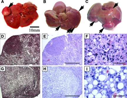
Prolonged consumption of high fat diet leads to dysplastic tumor formation. Dysplastic tumor nodules were identified in high fat diet livers as early as 9 months on diet (A: 1/2 mice). Gross images of livers maintained on a high fat diet for 14 months (B: 3/3 mice), and 20 months (C: 3/3 mice) respectively display multiple dysplastic nodules visible on the liver surface while no dysplasias were detected in mice maintained on regular diet for 9 or 14 months (0/3, 0/2 mice), even after microscopic sectional analysis. Histological analysis of liver sections revealed two different types of dysplastic nodules as either solid and non-fatty (D, E, F) or as tumors containing large droplets of lipid (G, H, I). Histological sections were stained with trichrome (D, G), as well as H&E (E, F, H, I) for analysis. High power magnification of H&E stained sections shows the cellular context of the two types of tumors (F, I). Scale bar = 10 mm (A, B, C); 1 mm (D, E, G, H); and 50 μm in (F, I).

