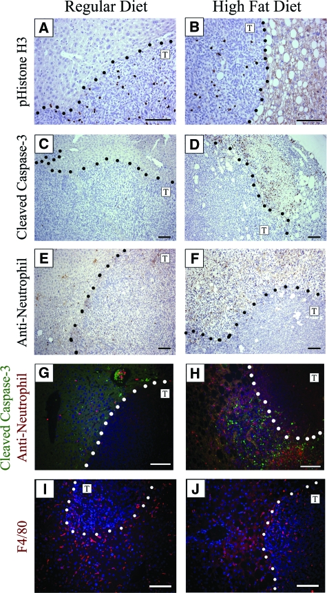
Figure 5.
Cellular responses to metastatic tumors in livers of mice maintained on regular diet or high fat diet. Dotted lines represent the tumor/liver boundary and the T designates the tumor side of the interface. No difference in cellular proliferation was detected from the staining index when comparing MC38 metastatic tumors or surrounding liver tissue using a phospho-histone H3 (pHistone-H3) antibody (A, B). Tumors contained very low numbers of apoptotic cells regardless of diet (C, D), while focal areas adjacent to MC38 induced tumors in the livers of 6/6 mice maintained on the high fat diet were found to contain high levels of cleaved caspase-3 staining indicating increased apoptosis (D). These densely apoptotic areas seen in the steatotic livers also contain high levels of inflammatory cells, demonstrated with anti-neutrophil staining (F), while neutrophils in regular diet livers were sparse (E). Co-immunofluorescence microscopy of cleaved caspase-3 (green) and anti-neutrophil (red) shows sparse staining near tumors in the liver of a mouse maintained on the regular diet (G), whereas mice maintained on high fat diet show high levels of distinct areas of peri-tumoral staining for both antibodies (H). Macrophages were found associated with the surrounding host tissue as well as within the tumor nodules in both sets of mice after staining with anti-F4/80 antibodies (red) and Hoechst for nuclei (blue) (I, J). Scale bar =50 μm (C, D, E, F) and 100 μm (A, B, G, H, I, J).

