Figure 2.
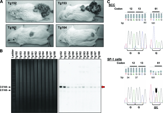
Analysis of transgene copy number and H-ras mutations in the K5.RasGRP1 mice and skin tumors. A: Photographs of transgenic tumor-bearing mice taken 10 weeks after a full-thickness incision wound was performed on the dorsal skin. Note one animal group with large tumors (top), and the other group with one small dorsal tumor only (bottom). B: Southern blot of NheI/BamHI digested genomic DNA from the K5.RasGRP1 mice subjected to the wounding protocol. The left panel corresponds to an ethidium bromide-stained agarose gel, showing equal DNA loading of the lanes. The right panel is the Southern blot autoradiogram from the same gel, obtained with a probe specific for detection of the Rasgrp1 transgene, and used to determined copy number. The red arrow points at the band containing the transgene (∼2.9 kb). C: H-ras sequencing analysis of genomic DNA obtained from K5.RasGRP1-derived tumors. A representative chromatogram is shown for DNA from SCC. Sequencing results for DNA from SP-1 cells are also shown as a positive control for H-ras mutations (black arrow). The corresponding amino acids for each codon are indicated below the chromatograms.

