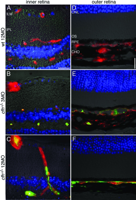
Figure 2.
Deposition of C3 along endothelial surfaces in the retina increases with age in the CFH-deficient mouse. Representative micrographs showing immunohistochemical colocalization of C3 deposition (green) and isolectin B4, a vascular endothelium marker (red) in retinae from 3-month-old cfh−/−(n = 5), 12-month-old cfh−/− (n = 5), and 12-month-old wild-type control mice (n = 5) sections. C3 deposition is absent in the inner (A) and outer (D) retina in the aged wild-type mouse. In the 3-month-old cfh−/− mouse, C3 protein is only detected on the endothelium of the deep microcapillaries in the inner retina outer plexus (B); it is also fragmentally deposited in the apical choroid (E). However, in the 1-year-old cfh−/− mouse, C3 has accumulated in the majority of endothelial vessel walls in the inner retina (C). It has also accumulated along the basal side of the RPE (F)—where the Choroidal vasculature has withered away as evidenced by the absence of isolectin-stained endothelium from large parts of the choroid. Inner limiting membrane, ILM; inner plexiform layer, IPL; and inner nuclear layer, INL. Scale bar = 20 μm.

