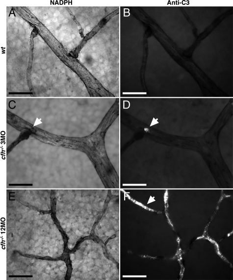
Figure 4.
Representative details of vasculature from primary plexus of flat mounted neural retina in 3-month-old (n = 3) and 1-year-old (n = 3) CFH deficient mice and 1-year-old control animals (n = 3) show increased deposition of C3 with age in knockout retina compared with the wild-type. Details of vessels stained with NADPH show the extent of vascular withering in the aged CFH-deficient retina (E) compared with those of young cfh−/− (C) and aged control retinae (A). C3-labeling (B, D, and F) exhibited extensive C3 accumulation in the aged cfh−/− (arrow in F) compared with young knockouts (D) and aged control animals (B). The vascular bed in the 3-month-old cfh−/− mouse retina appears relatively intact, however small ruptures at branching points (arrows in C and D) show small depositions of complement C3. The vascular bed in 1-year-old cfh−/− mice retina has a withered appearance (E) and around 80% of it is complement C3+ (F). Scale bar = 20 μm.

