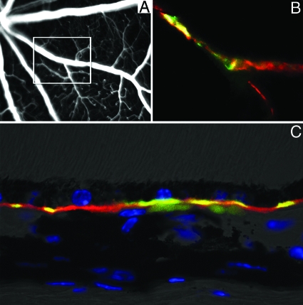
Figure 5.
Vascular depositions of activated C3 (C3b) fragments are present both in the inner retinal vasculature and along the Bruch’s membrane in the aged CFH-deficient mouse. High-resolution fluorescein angiogram shows venous beading in a 12-month-old cfh−/− (A). Detail of flat-mounted retina imaged in (A) show colocalized deposition of C3 (red) and C3b (green) (B) around vascular beading. Colocalization of inactive and active (C3b) forms of C3 along the Bruch’s membrane in the outer retina (C).

