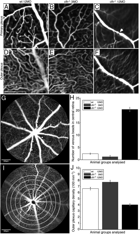
Figure 6.
Fluorescein angiographs illustrate vascular abnormalities in the aged CFH-deficient mouse retina compared with the age-matched control and the 3-month-old cfh−/− retina. A–F show high-resolution cSLO images of retinal vasculature at the level of the primary plexus (A–C) and at the level of the outer plexus (D–F) in 12-month-old control mice (A and D) (n = 5), 3-month-old cfh/ mice (B and E) (n = 4) and 1-year-old cfh/ mice (C and F) (n = 6). Vascular plexuses appear normal in the retina of aged control and 3-month-old cfh−/− mice. However, there is a large presence of venous beading in the primary plexus (arrow in C) and reduced retinal perfusion in the outer plexus in the retinae of 1-year-old cfh−/− mice. Scale bar = 100 μm. Quantification of vascular abnormalities in these animal groups revealed significant changes in the aged cfh−/− mouse compared with younger mutants and aged control mice. Venous beads (arrowheads) from a 1-year-old cfh−/− retina (G) in the primary plexus are defined as focal constrictions where the venous diameter is reduced by >40%. One-way analysis of variance analysis showed significantly larger number of venous beads in the primary plexus of the aged cfh−/− compared with the other two groups (P < 0.0001) H: Vascular perfusion in the outer plexus was quantified using a simple edge detection algorithm, which was applied radially at 100-μm intervals from the optic disk (as indicated by the concentric circles) I: Vascular density per 100 μm in the outer plexus was found to be significantly lower in the 1-year-old cfh−/− compared with that of the 3-month-old cfh−/− and 1-year-old control mice using one-way analysis of variance analysis (P < 0.001) (J).

