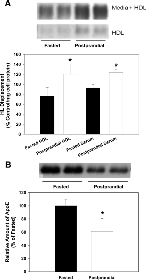
Figure 1.
Postprandial lipemia affects HL displacement by HDL. CHO-hHL cells were seeded in 6-well plates and treated with serum and HDL isolated from normolipidemic subjects for 45 minutes. The blood samples were taken after fasting, as well as 4 hours postprandial. The amount of HL present in HDL and media samples was measured using an immunochemical analysis. A: HL Western blot images are shown for fasted and postprandial HDL samples and also CHO-hHL media samples, after incubation with HDL (upper panel). HL displacement from the cell surface by HDL and serum was analyzed immunochemically and quantified by densitometry (lower panel). Averaged HL values are shown for serum and HDL samples obtained from three subjects after fasting and 4 hours postprandial, following an isocaloric meal. All values were calculated as a percentage of the HL displaced by the apoA-I control and were normalized to total cell protein. Values are the mean ± SD of triplicate determinations from one assay and represent three displacement experiments. Significance of difference relative to fasted sample *P < 0.01. B: The amount of apoE present in fasted and postprandial HDL samples was measured immunochemically. Western blot images are shown for the apoE in fasted and postprandial HDL samples (upper panel). HDL-apoE was quantified by densitometry and is shown (lower panel) relative to fasted values. Significant difference relative to fasted HDL, *P < 0.01.

