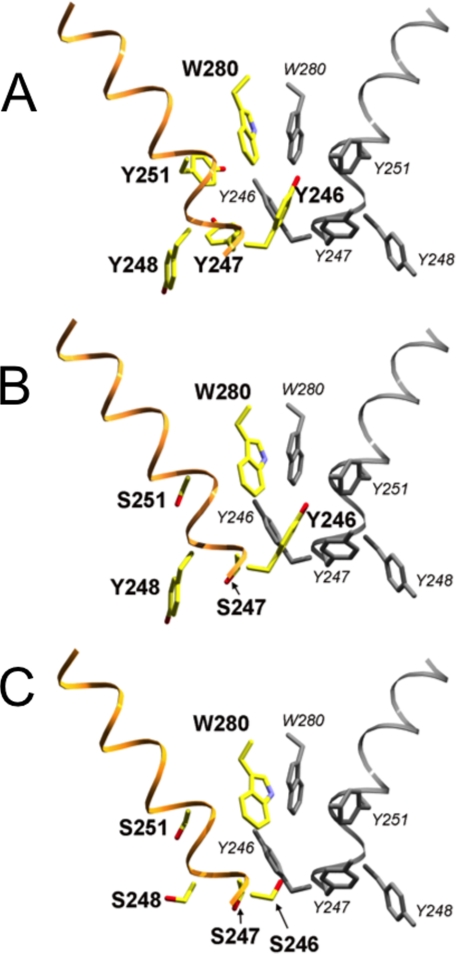
FIGURE 10.
Location of tyrosine → serine substitutions in B. thuringiensis PI-PLC mutants relative to the crystallographic dimer interface observed in the W47A/W242A mutant (PDB code 2OR2). Backbone ribbon representation with key side chains is shown in stick representation. A, crystallographic dimer (2OR2), with the second subunit shown in gray. Y247S/Y251S (B) and Y246S/Y247S/Y248S/Y251S (C) mutants, shown in Corey-Pauling-Koltun coloration, have been superimposed onto the first subunit of the A dimer structure; the second subunit from A is included to complete the hypothetical dimer interface. This comparison supports the hypothesis that the more active Y247S/Y251S mutant, but not the catalytically impaired Y246S/Y247S/Y248S/Y251S mutant, retains the key tyrosine residues needed for dimer formation.

