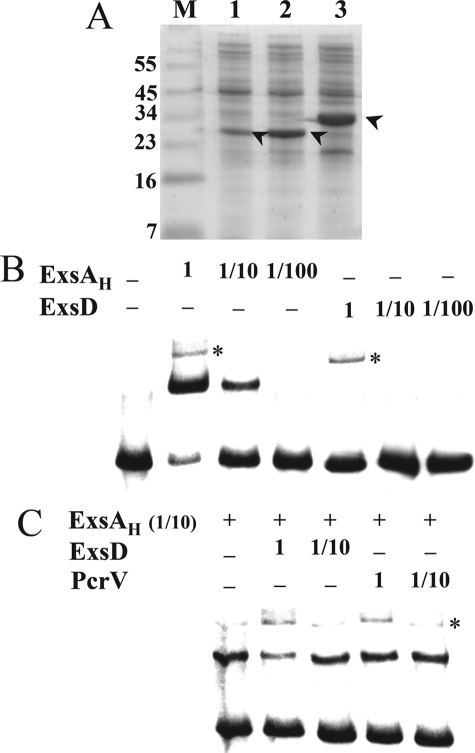
FIGURE 2.
ExsD interferes with ExsA binding to DNA. A, cytosolic extracts of cells producing ExsAH (lane 1), ExsD (lane 2), and PcrV (lane 3) were separated by SDS-PAGE and stained with Coomassie Blue. The bands corresponding to the proteins are indicated by an arrowhead. M, protein marker (in kDa). B, EMSA of the pC promoter with ExsAH and ExsD extracts. Biotinylated pC fragment (60-mer, 0.2 nm) was incubated for 15 min at 25 °C in the absence (−) or in the presence of 1 μl of indicated cytosolic fraction, either undiluted (0.7 mg/ml) (lane 1), diluted 10 (lane 1/10), or 100 times (lane 1/100). The samples were then electrophoresed, electrotransferred onto a nylon membrane, and revealed using Lightshift® chemiluminescent EMSA kit. C, 1 μl of ExsAH-cytosolic fraction, 10-fold diluted, was incubated with 1 μl of the indicated fractions (undiluted or diluted 10 times) 15 min prior to addition of pC probe. A nonspecific band (*) is revealed at the highest concentration of all extracts.

