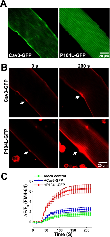
FIGURE 5.
Dominant effect of P104L-Cav3 produces defective membrane repair in native skeletal muscle. A, FDB muscle fibers from WT mice were transfected by in vivo electroporation to allow for transient expression of Cav3-GFP (left) and P104L-GFP (right) that were visualized by confocal microscopy. Confocal images showed that Cav3-GFP mainly targets to the sarcolemmal membrane, whereas P104L-GFP displayed a pattern indicative of intracellular retention of the mutant Cav3 protein. B, measurement of FM4-64 entry revealed severe defects in membrane repair capacity in fibers expressing P104L-GFP (lower), where excessive FM4-64 dye entry is observed following UV laser wounding (arrows) when compared with Cav3-GFP-expressing fibers (upper). C, quantitative assay of FM4-64 dye entry into skeletal muscle transiently expressing Cav3-GFP (blue trace) or P104L-GFP (red trace) or untransfected fibers on the same dish (Mock control, green trace). Data are means ± S.E. (error bars) for n = 18 fibers for each group from 3 independent electroporation with WT mice.

