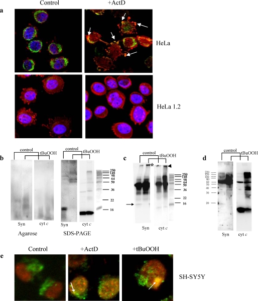
FIGURE 6.
Detection of inclusions and co-localization of hetero-polymerized Syn and cyt c aggregates in HeLa and SH-SY5Y cells after exposure to ActD or t-BuOOH. a, immunostaining of cyt c (green) and Syn (red) in intact HeLa cells and cyt c-deficient HeLa 1.2 cells. In control cells, cyt c appears in a punctate, perinuclear pattern of staining with little overlap with cytosolic Syn staining. ActD induces cytoplasmic blebbing, nuclear fragmentation, and transition from perinuclear to a diffuse staining pattern for cyt c. Focal regions of accentuated Syn staining, suggestive of aggregation, are observed, and this frequently co-localizes with focal regions of accentuated cyt c staining in cells with both preserved and fragmented nuclear contours. HeLa 1.2 clones engineered to express decreased levels of cyt c show a similar distribution of cyt c and Syn, although there is less cyt c immunoreactivity. HeLa 1.2 cells show preserved perinuclear cyt c and reduced propensity for clumped or aggregated Syn in response to the same doses of ActD, and there is no evidence of Syn and cyt c co-localization in these cells. b, Western blot analysis of Syn-cyt c aggregates after native agarose gel electrophoresis (left panel) and SDS-PAGE (right panel) in HeLa cells exposed to t-BuOOH. Left panel, staining with anti-Syn (Syn) and anti-cyt c antibodies (cyt c) reveals the appearance of co-localized hetero-oligomers after treatment of cells with t-BuOOH. Right panel, staining with anti-Syn (Syn) and anti-cyt c antibodies (cyt c) demonstrates disappearance of monomeric form of Syn, and accumulation of very high molecular weight Syn aggregates partially overlapping with aggregated forms of cyt c. Typical gels representative of three experiments are shown. c, detection of Syn-cyt c hetero-oligomers in HeLa cells exposed to t-BuOOH using immunoprecipitation with anti-Syn antibodies and Western blot analysis with anti-Syn and anti-cyt c antibodies after SDS-PAGE. Note that proteins immunoprecipitated with anti-Syn antibody contained high molecular weight aggregates positive for both Syn (asterisk) and cyt c (arrowhead) after treatment with t-BuOOH. Bands around 50 kDa likely correspond to heavy chains of immunoglobulins remaining after immunoprecipitation. Arrow indicates Syn monomer. Typical gels representative of three experiments are shown. d, Western blot analysis of Syn-cyt c aggregates after SDS-PAGE in mitochondria t-BuOOH from HeLa cells pretreated with t-BuOOH. Staining with anti-Syn (Syn) and anti-cyt c antibodies (cyt c) demonstrates accumulation of very high molecular weight Syn aggregates partially overlapping with aggregated forms of cyt c. Note that mitochondria from control cells contain significant amounts of monomeric cyt c but hardly detectable levels of monomeric Syn. After treatment with t-BuOOH, polymerized forms of Syn and cyt c are readily detectable, and the monomeric form of cyt c is decreased. Typical gels representative of three experiments are shown. e, immunostaining of cyt c (green) and Syn (red) in SH-SY5Y cells. Note that both proapoptotic agents, t-BuOOH and ActD, caused the appearance of LB-like inclusions revealed as yellow color in merged images (arrows). In normal SH-SY5Y cells, cyt c had a punctate mitochondrial distribution, and Syn was localized in the cytoplasm and perinuclear area. Following ActD or t-BuOOH treatment, Syn displayed aggregated pattern of distribution and overlapped with anti-cyt c staining (yellow spots on merged images).

