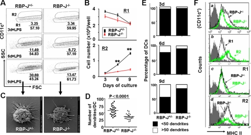
FIGURE 1.
RBP-J-deficient DCs showed maturation defects upon LPS stimulation. A, cells (2 × 10⁶) were obtained from BM and cultured in 24-well plates in the presence of GM-CSF and IL-4. Aliquots of cells were stimulated with LPS for 12 h on day 3, 6, and 9 of the culture, and were analyzed by FACS. B, number of CD11c+ DCs with low SSC (R1) and high SSC (R2). The number of cells per well in R1 and R2 in A was calculated and compared between RBP-J knockout and control mice. C, typical appearance of DCs from RBP-J knockout and control mice under an SEM. D, comparison of dendrite number between DCs from RBP-J knockout and control mice. DCs were cultured as above, and the number of dendrites of each DC was counted under an SEM. The average dendrite number was compared between RBP-J knockout and control DCs (n = 25). E, RBP-J knockout and control cells were cultured as in Fig. 1A. DCs with >50 dendrites and DCs with <50 dendrites were counted under an SEM. F, LPS-stimulated DCs were analyzed by FACS using anti-MHC II. The result represents three independent experiments. Bars represent means ± S.D. (n = 5). **, p < 0.01.

