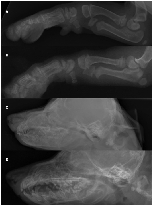
Figure 1. Radiographs of an OI affected and a control Dachshund demonstrating generalized osteopenia in canine OI.
(A) Foreleg of an affected Dachshund. Note the overall decreased radiopacity of the skeleton with the thin compact bone and inhomogeneous, shallow trabeculation in the entire foreleg. No pathologic fractures were seen in this puppy. (B) Foreleg of a control Dachshund. (C) Skull of an affected puppy. There is decreased opacity and poor delineation of the skull. Note the lack of visualization of the lamina dura of the dental alveoli leading to a “floating” appearance of the teeth, which themselves show a lack of mineralization. (D) Skull of a control dog.

