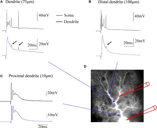
Figure 1.
Climbing fiber responses monitored by simultaneous double patch-clamp recordings from the soma and dendrites of cerebellar Purkinje cells. (A) Paired recordings from the soma (black trace) and the dendrite (blue trace). The dendritic patch electrode was located at about 75 μm distance from the soma. (B) In this recording, which was obtained from a different Purkinje cell, a more distal location (ca. 100 μm) was selected for the dendritic patch electrode. Small arrows in (A) and (B) point towards small, delayed spikelets seen in the dendritic recordings, that likely reflect sodium action potentials generated in the axon (see corresponding, but larger spikes in the somatic recordings), which passively spread into the dendrite. (C) At a far more proximal location (ca. 10 μm), the dendritic recording resembles the complex spike recorded in the soma, but already at this distance an attenuation of the initial, fast sodium spike can be seen. (D) Recording configuration of the experiments. This picture was assembled for illustration purposes only. The recordings were obtained from cerebellar slices prepared from P28-35 Sprague-Dawley rats. Slices were perfused with standard ACSF (as described in Coesmans et al., 2004) bubbled with 95% O2 and 5% CO2. The recordings were performed at near-physiological temperature (31–34°C) using an EPC-10 amplifier (HEKA Electronics, Germany). Currents were filtered at 3 kHz, digitized at 10 kHz, and acquired using Fitmaster software. The recording electrodes were filled with a solution containing (in mM): 9 KCl, 10 KOH, 120 K gluconate, 3.48 MgCl2, 10 HEPES, 4 NaCl, 4 Na2ATP, 0.4 Na3GTP, and 17.5 sucrose (pH 7.25). Patch electrodes used for somatic recordings had electrode resistances of 3–4 MΩ, and patch electrodes used for dendritic recordings had electrode resistances of 7–10 MΩ. For climbing fiber stimulation, glass pipettes were used that were filled with ACSF.

