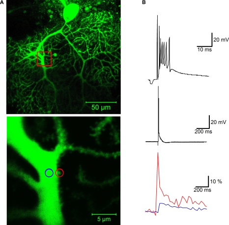
Figure 2.
Calcium signaling in a dendritic spine evoked by climbing fiber activity. (A) Top: A rat Purkinje cell (P36) was filled through the patch pipette with the fluorescent calcium indicator Oregon Green BAPTA-2 (200 μM). The red box indicates the magnification area. Calcium transients were recorded in a spine (red circle) and in the shaft (blue circle) of the primary dendrite using a Zeiss LSM 5 Exciter confocal microscope and a ×63 Zeiss Apochromat objective (bottom picture). (B) Climbing fiber stimulation evokes a complex spike shown at two different time scales (top), which is associated with a large calcium transient monitored in the spine (red trace), and a smaller transient in the shaft (blue trace; bottom). The calcium traces represent averages of 10 transients (acquired at 30 s delays), and are displayed as normalized fluorescence changes (ΔF/F). For image acquisition and analysis we used ZEN software (Carl Zeiss MicroImaging, Thornwood, NY, USA). Patch-clamp recording details are described in Figure 1.

