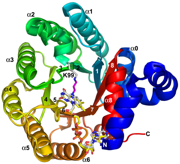
Figure 1.
Superposition of N-propargylglycine-inactivated TtPRODH and oxidized TtPRODH. The proteins are colored using a rainbow scheme, with blue at the N-terminus and red at the C-terminus. Strands and helices of the (βα)8 barrel are labeled. The FAD cofactors are colored white in the inactivated enzyme and yellow in the oxidized enzyme. Lys99 of the inactivated enzyme is represented in white sticks. The three-carbon link between the Lys99 ε-amino group and the FAD N5 is shown in magenta. This figure and others were prepared with PyMOL (W. L. DeLano (2002) The PyMOL Molecular Graphics System).

