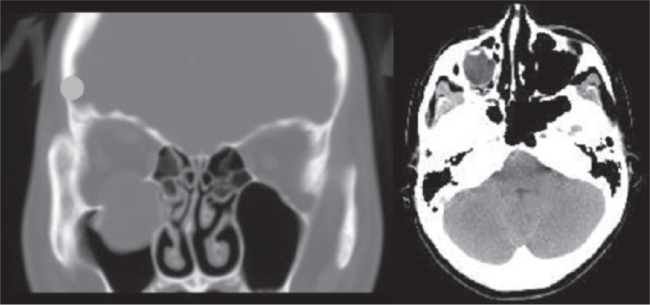
Figure 1.
Computed tomographic images showing blow-out fracture of the lower right orbital wall. Left: Coronal section showing that the right eyeball is not perforated but has shifted downward into the maxillary sinus through the fracture of the lower orbital wall. right: Axial section showing that her right eyeball was displaced.

